Visio-Vestibular Markers for Concussion
There is significant evidence that concussion can lead to visio-vestibular deficits post-concussion such as impaired eye movement and pupil response. Over a series of research publications, CIRP’s Minds Matter researchers describe these deficits as potential objective markers seen in the visual system after concussion and their association with prolonged recovery.
One of the primary tools Minds Matter clinicians have to assess these visual deficits is a visio-vestibular exam (VVE) advanced at CHOP. Researchers have been testing the utility of the VVE to detect deficits after concussion in pediatric and adolescent patients and the use of eye tracking and pupillometry technology to objectively measure these deficits. They have learned that such clinical tools are both effective and feasible and important for diagnosis and recovery.
This line of research has important implications for the management of visual deficits after concussion. Active rehabilitation therapy can be used to treat those with prolonged persisting symptoms, and specific academic accommodations targeting these visio-vestibular deficits can be made for these children when returning to the learning setting.
Research Projects
- Visio-Vestibular Deficits Post-Concussion
Vision and Vestibular System Dysfunction Predicts Prolonged Concussion Recovery in Children
This study looked retrospectively at 432 randomly selected children ages 5-18 years who were seen in a specialty concussion program, of which 88 percent presented with a visio-vestibular deficit on initial clinical examination. Deficits in smooth pursuit and vestibular-ocular reflex function, accommodative amplitude and balance predicted prolonged concussion recovery.
Near Point of Convergence After Concussion in Children
In a study of 275 patients age 5-18 with concussion, nearly one-fourth were found to have abnormal near point of convergence (NPC) on physical exam. About half recovered with standard clinical care over a median of 4.5 weeks, 40% recovered a median of 11 weeks post-injury with vestibular therapy and the remainder had persistent abdominal NPC that necessitated referral to a developmental optometrist for vision therapy.
Vision Diagnoses Are Common After Concussion in Adolescents
In a single-center cross-sectional study of 100 patients with concussion enrolled at the Minds Matter concussion program at CHOP, 69 percent had at least one vision diagnosis after concussion. Clinician researchers found the Convergence Insuffiency Symptom Survey (CISS) to be a highly cost-effective means for general practitioners to identify patients with concussion-related vision deficits that could require referral to concussion specialist or eye care professional.
Vestibular Deficits Following Youth Concussion
In a retrospective cohort study of patients ages 5 to 18 referred to a sports medicine clinic, researchers sought to characterize the prevalence and recovery of pediatric patients with concussion who manifest clinical vestibular deficits, as well as describe the correlation of vestibular deficits with neurocognitive function based on computerized neurocognitive testing. Vestibular deficits were highly prevalent, associated with longer recovery and poorer outcomes in neurocognitive testing.
Oculomotor and Cognitive Test in Youth with Concussion
This study looked for correlations between different baseline assessments for concussion used for child and adolescent athletes: King-Devick (K-D) is a number-naming test assessing saccadic eye movement, ImPACT assesses working memory, visual motor speed and reaction time, while SCAT3 assesses working memory, concentration, and balance. In addition to these assessment tools, researchers wanted to see if useful information could be also obtained by measuring near point of convergence (NPC), the point at which a single visual object becomes double in near vision. In this study of youth hockey players ages 6-18 years, researchers ultimately determined that use of multiple assessment tools in a clinical evaluation of pediatric concussion is warranted rather than relying on any single tool. Also, the results suggest that the pediatric population may require more frequent than annual baseline testing. Additional research is needed to determine which combination of assessments will be most useful and non-overlapping.
Frequency of oculomotor disorders in adolescents 11 to 17 years of age with concussion, 4 to 12 weeks post injury
Trajectories of Visual and Vestibular Markers of Youth Concussion
Developmental Effects on Pattern Visual Evoked Potentials Characterized by Principal Component Analysis
Relationship between Visually Evoked Effects and Concussion in Youth
Disparity vergence differences between typically occurring and concussion-related convergence insufficiency pediatric patients
- Visio-Vestibular Exam
Evaluating Visio-Vestibular Exam (VVE)
The visio-vestibular exam (VVE) identifies visual and vestibular system deficits which aid in the diagnosis of concussion and can predict prolonged recovery in those who are identified as having such deficits. These systems are responsible for integrating balance, vision and movement. Understanding these deficits early can have a substantial impact on a child’s visual function ability to return to learn and sports after a concussion.
The VVE screening assesses how eyes track a moving object, jump quickly between visual targets and their ability to view an object at near distance without double vision. Taken in the context of a preceding injury, abnormalities on this testing can assist in the diagnosis of concussion, as well as potentially aid in predicting children who will suffer from prolonged symptoms.
The Consensus Statement on Concussion in Sport, published by the Concussion in Sport Group, endorses the VVE as part of the recommended best practice for the Child Sports Concussion Office Assessment Tool - Child SCOAT6 tool.
Use of the Vestibular and Oculomotor Examination For Concussion In a Pediatric Emergency Department
This study analyzed patient charts of 400 children ages 6-18 who presented to the emergency department (ED) with head injury and found that 64 percent of the patients overall were assessed using the VVE. Of those ultimately diagnosed with concussion, 73 percent were assessed using the VVE. Nine percent of patients diagnosed with a concussion had one or fewer symptoms but abnormal exam findings, highlighting that the VVE can assist in more accurately identifying concussed patients. The VVE was least likely to be performed in those children with non-sports related injuries, minimal concussion symptoms, and no prior history of concussion. With training and clinical support tools, pediatricians, emergency medicine clinicians, and advanced practice practitioners are able to conduct the VVE in a high volume acute care setting.
Watch Dr. Daniel Corwin demonstrate how to administer the elements of the Visio-Vestibular Examination for Concussion:
Reliability of the visio-vestibular examination for concussion among providers in a pediatric emergency department
Characteristics and Outcomes for Delayed Diagnosis of Concussion in Pediatric Patients Presenting to the Emergency Department
Visio-Vestibular Deficits in Healthy Child and Adolescent Athletes
Pre- and post-season visio-vestibular function in healthy adolescent athletes
- Pupillary Light Reflex (PLR)
The Utility of Pupillary Light Reflex Metrics as a Physiologic Biomarker for Adolescent Sport Related Concussion
The pupil’s response to light is an autonomic function with metrics easily captured via an automated dynamic infrared pupillometry hand-held device. In this study, researchers explored whether PLR could distinguish between concussed and non-concussed adolescents. Researchers were interested in nine specific PLR metrics that objectively assess autonomic function. In a prospective cohort of adolescent athletes (ages 12-18) that included both healthy athletes (n = 134) and athletes with a diagnosis of sport-related concussion (SRC; n = 98), they found significant differences between concussed athletes and healthy adolescents for eight of the nine PLR metrics. They also observed differences for females with concussion.
Watch Dr. Christina Master explain the novel use of a pupillometer device to diagnose and monitor concussions:
- Automated Eye-Tracking Assessment
Evaluating Automated Eye-Tracking Assessment
The automated eye tracking assessment is a rapid, objective, non-invasive aid in the diagnosis of concussion and it does not require an individual patient’s pre-injury baseline as a comparison to identify a concussion. This eye-tracking methodology reflects natural automatic physiologic brain activity and, thus, can be an objective alternative to traditional subjective, symptom-based assessments.
During the assessment, a clinician records a patient’s binocular eye movements with an SR Research Eyelink 1000 eye tracker while they are watching a 220-second video move continuously within a square aperture on the monitor as the patients head is stabilized on a chin rest to minimize head movement. Data collected are automatically processed using Oculogica software to yield metrics relevant to conjugacy, or the ability of the eyes to move well together. In collaboration with other institutions, CIRP researchers evaluated this technology as an objective method for identifying concussions in young patients. In 2019, the Oculogica EyeBox received FDA authorization as one of the few devices cleared for commercial use in objective diagnosis of concussion.
Eye Tracking Metrics Differences among Uninjured Adolescents and Those with Acute or Persistent Post-Concussion Symptoms
Objective Eye Tracking Deficits Following Concussion for Youth Seen in a Sports Medicine Setting
This study evaluated eye tracking measurements among adolescents within 10 days of the injury and healthy controls (n = 79). From among 17 metrics analyzed in this study, one metric measured by the automated eye tracking assessment stood out: The right eye’s ability to track an object predicted overall symptom severity among patients with concussion as compared to healthy controls.
Eye Tracking as a Biomarker for Concussion in Children
This study evaluated eye tracking measurements among children with concussion seen in a concussion referral setting with a mean of 22 days out from injury and healthy controls (n = 139). For these patients there were 12 metrics from 89 evaluated that were significantly different between concussed and non-concussed children. The assessment’s detection of concussion was independent from the number of symptoms reported by the patient.
Reliability of Objective Eye-Tracking Measures Among Healthy Adolescent Athletes
This study evaluated healthy young athletes across two different testing sessions to evaluate the reliability of objective eye tracking metrics. No significant differences in outcomes were found in 13 eye movement variables across the two testing times. These results suggest that these automated and quantitative eye movement metrics are relatively stable among a group of healthy youth athletes.
- Evaluating Measures of Gait and Balance
Prognosis for Persistent Post Concussion Symptoms using a Multifaceted Objective Gait and Balance Assessment Approach
A prospective cohort study of youth and young adult athletes determined that quantitative dual-task gait measures provide useful information for PPCS prognosis. Single-task gait, stance, and cognitive performance were not associated with PPCS. Researchers recommend that independent validation should be done in larger cohorts to determine utility.
Clinical and Device-based Metrics of Gait and Balance in Diagnosing Youth Concussion
This study of 171 subjects (81 concussed, 90 non-concussed) compared the discriminatory ability of three measures of gait and balance: a device-based measure (a biomechanical force plate device) and two clinical measures (the modified balance error scoring system, or mBESS, of the Sport Concussion Assessment Tool, and the complex tandem gait used in the VVE). Investigators found that each test possessed moderate discriminatory ability, with the two clinical measures performing as well as, if not slightly better than, the device-based measure. In addition, investigators found the complex tandem gait possessed components (having the subject walk backward with his or her eyes closed) with the highest sensitivity for ruling out concussion.
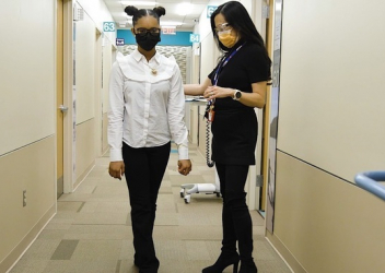
Watch Dr. Daniel Corwin demonstrate how to administer Complex Tandem Gait, an evaluation of gait to diagnose a concussion:
- Evaluating Saccades and Gaze Stability Assessments
Assessment of Saccades and Gaze Stability in the Diagnosis of Pediatric Concussion
A cohort study of 69 concussed athletes and 69 healthy athletes evaluated the discriminatory ability of saccades and gaze stability testing at different repetition increments to determine the optimal number of repetitions to differentiate youth athletes with and without concussions. This study of showed that increasing the number of repetitions from 10 to 20 significantly increased the sensitivity of the test. When combining the four tests (saccades and gaze stability in both the horizontal and vertical plane), the Minds Matter Concussion team found discriminatory ability was maximized with a cut-off of 20 repetitions. The findings in this study suggest that a higher threshold level of repetitions of two commonly used visio-vestibular assessments enables clinicians to more accurately diagnose youth concussion.


